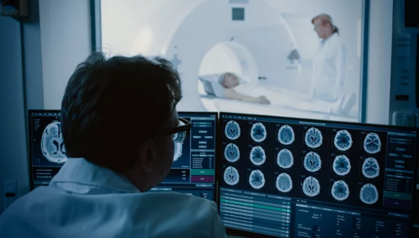
Magnetic Resonance Imaging (MRI) is one of the most advanced diagnostic tools available for cancer detection. With its ability to capture high-resolution images of soft tissues, MRI often identifies tumors earlier than traditional imaging methods. However, its high sensitivity can also be a drawback—leading to false positives. A false positive occurs when a scan suggests the presence of cancer, but further testing reveals no malignancy.
False positives in cancer MRI screening can cause unnecessary anxiety, invasive procedures, and increased healthcare costs. This article explores why false positives happen, their impact on patients, and the best practices for reducing them without compromising the ability to detect cancers early.
Why False Positives Occur in MRI Cancer Screening
MRI is designed to be extremely sensitive, which means it can detect even the smallest irregularities. But not all abnormalities are cancer. Common reasons for false positives include:
- Benign conditions mimicking cancer (fibroadenomas in the breast, benign prostatic hyperplasia in the prostate).
- Normal anatomical variations that appear suspicious.
- Post-treatment changes such as scar tissue or inflammation mistaken for recurrence.
- Technical factors like patient movement or poor image quality.
- Overinterpretation when radiologists err on the side of caution to avoid missing a true cancer.
The Impact of False Positives on Patients and Healthcare Systems
False positives are not just statistical errors—they have real consequences:
- Emotional distress: Patients may experience unnecessary fear and anxiety.
- Unnecessary procedures: Follow-up biopsies, surgeries, or additional imaging increase physical and emotional burdens.
- Financial costs: Extra tests raise healthcare expenses for patients and insurers.
- Overdiagnosis risk: Some conditions detected might never progress to clinical cancer, yet patients undergo treatment.
Reducing false positives is therefore essential for balancing early detection with patient safety.
Strategies for Reducing False Positives in MRI Cancer Screening
1. Standardized Imaging Protocols
Following evidence-based MRI protocols reduces variability in imaging and interpretation. Organizations like the American College of Radiology (ACR) have developed systems such as:
- BI-RADS for breast MRI.
- PI-RADS for prostate MRI.
- LI-RADS for liver MRI.
These structured frameworks help radiologists assess lesions consistently and reduce unnecessary recalls.
2. Use of Multi-Parametric MRI (mpMRI)
For cancers like prostate or breast, combining different MRI sequences (T2-weighted, diffusion-weighted imaging, and contrast-enhanced imaging) provides a more complete picture. This multi-sequence approach improves specificity, lowering the chance of false positives.
3. Incorporating Artificial Intelligence (AI) and Radiomics
AI-powered imaging analysis can identify subtle patterns and reduce human error. Radiomics—extracting quantitative features from MRI scans—provides additional data to distinguish between benign and malignant lesions more accurately.
4. Radiologist Training and Experience
Interpretation accuracy improves with experience. Specialized training in oncologic MRI helps radiologists recognize benign findings that mimic cancer, reducing overdiagnosis.
5. Comparative Imaging
Comparing new MRI scans with prior images helps identify stable, non-cancerous lesions and prevents unnecessary alarms.
6. Patient Selection for MRI Screening
Instead of using MRI for the general population, many public health programs reserve it for high-risk groups (e.g., women with BRCA mutations, men with elevated PSA and abnormal exams). Targeted screening reduces unnecessary testing and lowers the chance of false positives in low-risk individuals.
7. Second Opinions and Double Reading
In complex cases, involving two radiologists—or using tele-radiology for second opinions—can reduce interpretation errors and unnecessary biopsies.
Examples in Specific Cancers
Breast Cancer
Breast MRI is highly sensitive, especially for women with dense breast tissue. However, false positives are common. Studies show that using BI-RADS scoring and combining MRI with mammography improves specificity.
Prostate Cancer
Multi-parametric MRI is now a standard in prostate cancer evaluation. It significantly reduces unnecessary biopsies when used alongside PSA testing.
Liver Cancer
For patients with chronic liver disease, MRI with LI-RADS classification helps distinguish between benign lesions and hepatocellular carcinoma, reducing misinterpretation.
Future Directions in Reducing False Positives
- AI-enhanced triage systems that automatically flag high-risk lesions while filtering benign findings.
- Machine learning algorithms that integrate patient data (genetics, biomarkers, history) with imaging for higher specificity.
- Faster, higher-resolution MRI machines that reduce artifacts and improve clarity.
- Global adoption of standardized reporting systems to ensure consistency across healthcare systems.
Conclusion: Striking the Right Balance
False positives in MRI cancer screening are a double-edged sword. While high sensitivity ensures early cancer detection, unnecessary recalls and interventions can harm patients emotionally, physically, and financially. By applying best practices—standardized protocols, advanced technologies, radiologist training, and careful patient selection—healthcare systems can reduce false positives while preserving MRI’s life-saving potential.
The future of MRI lies in precision: using the technology wisely, interpreting it accurately, and ensuring that every positive result truly guides patients toward better health outcomes.
Also Read :