Magnetic Resonance Imaging (MRI) has transformed the field of oncology by offering detailed, non-invasive imaging that helps detect, stage, and monitor cancer. Unlike traditional imaging techniques, MRI provides high-resolution visualization of soft tissues, making it invaluable in diagnosing cancers that might otherwise be missed. Real-world case studies demonstrate how MRI is applied across different cancer types, influencing treatment decisions and improving outcomes.
This article presents several case studies highlighting the role of MRI in cancer diagnosis and treatment, illustrating how this technology shapes patient care in practice.
Case Study 1: Breast Cancer Detection in High-Risk Patients
Background
A 42-year-old woman with a strong family history of breast cancer and a BRCA1 mutation underwent annual mammography screenings. While mammograms detected no abnormalities, she continued to experience concerns due to her genetic risk.
MRI Findings
Breast MRI revealed a small, enhancing lesion in the upper outer quadrant of the left breast. The lesion measured only 7 mm—too small to be clearly seen on mammography.
Outcome
A targeted biopsy confirmed early-stage invasive ductal carcinoma. Because of the early detection, the patient underwent lumpectomy followed by radiation, avoiding more aggressive systemic therapies.
Key Takeaway
MRI is particularly valuable for high-risk women, detecting cancers that mammograms may miss, especially in dense breast tissue.
Case Study 2: Prostate Cancer Evaluation With Multiparametric MRI
Background
A 60-year-old man presented with an elevated prostate-specific antigen (PSA) level but had inconclusive biopsy results. Traditional ultrasound did not provide sufficient clarity.
MRI Findings
Multiparametric prostate MRI (mpMRI) showed a suspicious lesion in the peripheral zone of the prostate with restricted diffusion and abnormal contrast enhancement. The lesion was scored as PI-RADS 4, indicating high suspicion of clinically significant cancer.
Outcome
MRI-targeted biopsy confirmed Gleason score 7 prostate cancer. Based on staging information, the patient was treated with a combination of radiation therapy and hormone therapy.
Key Takeaway
mpMRI improves diagnostic accuracy in prostate cancer, guiding targeted biopsies and reducing unnecessary procedures.
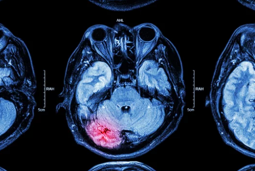
Case Study 3: Brain Tumor Characterization With Functional MRI
Background
A 35-year-old patient developed persistent headaches and seizures. A CT scan suggested a possible brain tumor but lacked detail about its boundaries.
MRI Findings
MRI identified a lesion in the left temporal lobe. Functional MRI (fMRI) was performed to map brain activity and assess areas critical for speech and motor function.
Outcome
The neurosurgical team used MRI and fMRI data to plan a tumor resection that removed the cancerous tissue while preserving language function. Postoperative recovery was smooth, and adjuvant therapy was initiated.
Key Takeaway
Functional MRI is essential in brain cancer management, guiding surgery to maximize tumor removal while protecting vital brain functions.
Case Study 4: Liver Cancer in Cirrhosis Patients
Background
A 58-year-old man with chronic hepatitis C and cirrhosis underwent routine ultrasound screening. A small lesion in the liver was detected but was indeterminate.
MRI Findings
Dynamic contrast-enhanced liver MRI with LI-RADS classification identified the lesion as LI-RADS 5—highly suggestive of hepatocellular carcinoma (HCC).
Outcome
The diagnosis was confirmed, and the patient underwent transarterial chemoembolization (TACE), followed by evaluation for liver transplantation.
Key Takeaway
MRI provides superior sensitivity and specificity in distinguishing between benign nodules and malignant liver lesions, crucial for patients with underlying liver disease.
Case Study 5: Monitoring Treatment Response in Bone Cancer
Background
A 16-year-old adolescent diagnosed with osteosarcoma of the femur began chemotherapy. Monitoring response was critical to adjust treatment and plan surgery.
MRI Findings
Baseline MRI showed extensive tumor involvement with surrounding soft tissue extension. After three cycles of chemotherapy, repeat MRI showed significant reduction in tumor size and necrosis within the lesion.
Outcome
The favorable response allowed the surgical team to perform limb-sparing surgery instead of amputation. The patient continued adjuvant chemotherapy with improved prognosis.
Key Takeaway
MRI plays a vital role in monitoring tumor response, directly influencing surgical decisions and long-term quality of life.
Lessons From These Case Studies
Across different cancers, MRI demonstrates its value in several ways:
- Early detection of small or hidden tumors.
- Improved specificity through multiparametric and contrast-enhanced imaging.
- Treatment planning by mapping tumor extent and functional impact.
- Monitoring therapy response, helping clinicians adjust strategies.
- Reducing unnecessary interventions by distinguishing between benign and malignant lesions.
The Future of MRI in Cancer Care
Advances in technology promise even greater impact:
- AI-enhanced interpretation to reduce false positives and improve diagnostic confidence.
- Radiomics to extract deeper insights from imaging beyond visual assessment.
- Molecular MRI techniques for detecting cancer at the cellular level.
- Global standardization of protocols to ensure uniform quality in patient care.
Conclusion: MRI as a Game-Changer in Oncology
MRI is more than just an imaging tool—it is a decision-making partner in cancer care. From detection in high-risk patients to guiding surgery and monitoring treatment response, MRI has proven to be indispensable. Real-world case studies illustrate how MRI continues to transform outcomes, offering precision and confidence in managing one of the most complex diseases of our time.
Also Read :