Cancer diagnosis and management rely heavily on advanced imaging techniques. Two of the most commonly used modalities are Magnetic Resonance Imaging (MRI) and Positron Emission Tomography (PET) scans. Both provide critical information about tumors, but they work differently, have distinct strengths, and are suited for different purposes. Understanding the differences can help patients and healthcare providers make informed decisions about diagnosis, staging, and treatment planning.
This article compares MRI and PET scans in cancer detection, exploring their advantages, limitations, and optimal clinical uses.
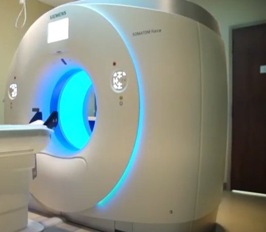
How MRI and PET Scans Work
Magnetic Resonance Imaging (MRI)
MRI uses strong magnetic fields and radio waves to create highly detailed images of organs, tissues, and tumors. It is particularly effective for:
- Soft tissue imaging (brain, breast, liver, prostate).
- Determining tumor size, structure, and location.
- Evaluating surrounding tissue involvement without radiation exposure.
MRI can also use contrast agents to enhance visualization and specialized sequences like diffusion-weighted imaging (DWI) to assess cellular density, helping differentiate benign from malignant lesions.
Positron Emission Tomography (PET) Scan
PET scans use small amounts of radioactive tracers, usually combined with a sugar molecule like FDG (fluorodeoxyglucose). Cancer cells, which metabolize sugar faster than normal cells, absorb the tracer and appear as bright “hot spots” on the scan. PET scans are particularly effective for:
- Detecting metastasis (cancer spread).
- Identifying active cancer tissue.
- Monitoring treatment response.
PET scans are often combined with CT (PET/CT) or MRI (PET/MRI) to provide both functional and structural information.
MRI Strengths in Cancer Detection
- High-resolution imaging: MRI provides detailed pictures of soft tissues, essential for tumors in the brain, breast, liver, and prostate.
- No radiation: Safe for repeated imaging, especially in children or patients needing long-term follow-up.
- Structural assessment: Excellent for surgical and radiation planning due to clear anatomical detail.
- Functional imaging options: Advanced MRI techniques (DWI, perfusion MRI) provide insights into tumor biology.
PET Scan Strengths in Cancer Detection
- Functional assessment: PET identifies active cancer cells based on metabolism, often detecting tumors before structural changes appear.
- Whole-body imaging: Effective for spotting metastases in multiple organs.
- Treatment monitoring: PET scans can evaluate early response to chemotherapy or targeted therapy.
- Complementary to structural imaging: PET combined with CT or MRI provides a comprehensive picture of both anatomy and cancer activity.
Limitations of MRI
- Limited functional information: MRI shows anatomy well but does not directly indicate metabolic activity.
- Less effective for small or diffuse metastases: Certain cancers like lung nodules may be better detected with CT or PET.
- Longer scan times: MRI can take 30–60 minutes, which may be difficult for some patients.
- Cost and availability: MRI is more expensive and less available in some regions.
Limitations of PET Scans
- Radiation exposure: Although relatively low, repeated PET scans involve exposure to radioactive tracers.
- Lower anatomical resolution: PET alone provides limited structural detail; combining with CT or MRI is usually required.
- False positives: Infections, inflammation, or benign conditions may appear as “hot spots,” leading to unnecessary follow-up tests.
- High cost: PET scans are expensive and not always covered for screening purposes.
When MRI Is Preferable
MRI is often the first choice in the following situations:
- Tumors in soft tissues such as the brain, breast, or prostate.
- Detailed anatomical evaluation for surgical or radiation planning.
- Pediatric patients or patients requiring frequent follow-up scans.
- High-resolution imaging for organ-specific cancers.
When PET Scans Are Preferable
PET scans are particularly useful for:
- Detecting metastatic cancer or recurrence.
- Evaluating tumor activity and aggressiveness.
- Monitoring response to chemotherapy or immunotherapy.
- Whole-body assessment in cancers with high metastatic potential (lung, lymphoma, melanoma).
Combining MRI and PET: The Best of Both Worlds
In many cases, PET/MRI or PET/CT combines the strengths of both modalities:
- Structural detail from MRI.
- Functional activity from PET.
- Improved accuracy in staging and treatment planning.
This hybrid approach is increasingly used in complex cancers, such as brain tumors, prostate cancer, and metastatic disease, providing a comprehensive understanding of both anatomy and tumor biology.
Key Takeaways
- MRI excels in anatomical detail and soft tissue imaging, ideal for localized tumor detection and planning interventions.
- PET excels in functional and metabolic imaging, ideal for detecting metastasis and monitoring treatment response.
- Neither MRI nor PET detects all cancers perfectly, and each has limitations depending on tumor type, size, and location.
- Hybrid imaging (PET/MRI or PET/CT) often offers the most complete evaluation, combining structural and functional information.
- The choice between MRI and PET should be guided by cancer type, stage, risk factors, and clinical goals.
Conclusion: Choosing the Right Imaging Tool
Both MRI and PET scans are indispensable in modern oncology, but they serve different purposes. MRI provides unparalleled anatomical detail, while PET highlights metabolic activity, making them complementary rather than competing technologies. Optimal cancer detection often involves a strategic combination of both, tailored to the patient’s needs.
By understanding the strengths and limitations of MRI and PET, clinicians can make informed decisions that enhance diagnosis, guide treatment, and improve patient outcomes.
Also Read :