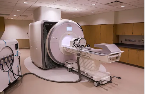
Getting an MRI can feel like a mysterious process. You lie in a narrow tube, listen to the rhythmic tapping of the machine, and hope for answers. But what happens after you leave the imaging center? Where do your scans go, who looks at them, and how do they influence your care? Understanding the journey of your MRI can demystify the process and show why this technology is so critical in cancer diagnosis and treatment.
Step 1: Image Capture and Processing
Once your MRI scan is complete, the machine generates raw data in the form of digital signals. These signals are processed by powerful computers to produce detailed cross-sectional images of your body.
Key points about this stage:
- High-resolution imaging: The computer transforms signals into high-definition images of soft tissues, organs, and blood vessels.
- Different sequences: MRI produces multiple types of images, each highlighting specific tissue properties, such as diffusion-weighted images or contrast-enhanced sequences.
- Data storage: The images are stored digitally in a secure system called a PACS (Picture Archiving and Communication System), allowing authorized clinicians to access them at any time.
This step ensures that your scans are accurate, detailed, and ready for interpretation.
Step 2: Radiologist Review
After processing, your MRI is reviewed by a radiologist, a medical doctor specializing in imaging interpretation. The radiologist examines every slice of your scan for abnormalities, paying attention to:
- Tumor presence and size: Detecting suspicious masses or lesions.
- Tissue characteristics: Differentiating between healthy tissue and possible malignancies.
- Changes over time: Comparing with previous scans to track growth or shrinkage.
- Functional information: Assessing blood flow, cellular density, and other features depending on the type of MRI sequence used.
Radiologists may also consult with colleagues, especially for complex cancer cases, ensuring the findings are accurate and actionable.
Step 3: Report Generation
After interpreting the images, the radiologist prepares a comprehensive report. This document is the primary communication tool between the radiologist and your treating physicians.
The report typically includes:
- Findings: A detailed description of any abnormal areas, their size, location, and characteristics.
- Impression: The radiologist’s overall assessment, including possible diagnoses.
- Recommendations: Suggested next steps, such as biopsy, additional imaging, or follow-up scans.
This report guides your oncologist or surgeon in making informed decisions about your care.
Step 4: Integration Into Your Medical Record
Your MRI images and the radiologist’s report are uploaded to your electronic medical record (EMR). This ensures that:
- Your care team has full access: Oncologists, surgeons, and primary care doctors can review your scans and reports.
- Continuity of care: Future imaging studies can be compared to track disease progression or response to treatment.
- Patient access: Many hospitals allow patients to view their MRI images and reports through secure online portals.
Integration into your medical record ensures that your MRI has a lasting impact on your diagnosis and treatment plan.
Step 5: Multidisciplinary Use
For cancer patients, MRI findings often feed into multidisciplinary care planning. Your images may be reviewed in a tumor board meeting, where specialists collaborate to determine the best course of action.
The MRI can influence:
- Surgical planning: Determining tumor location and margins.
- Radiation therapy: Mapping the tumor for precise targeting.
- Chemotherapy or targeted therapy: Assessing whether treatment is effective or needs adjustment.
- Follow-up schedules: Determining the frequency of future imaging based on risk and tumor behavior.
Step 6: Long-Term Storage and Research Use
Once your MRI is interpreted and integrated into your record, it is stored securely for the long term. Hospitals typically retain imaging for years, allowing:
- Future comparisons: Critical for detecting recurrence or monitoring chronic conditions.
- Research and teaching: De-identified images may contribute to studies that improve cancer detection and treatment methods.
- Quality assurance: Hospitals can review imaging to maintain diagnostic accuracy and standards.
This long-term storage ensures that your MRI continues to serve both your care and broader medical advancements.
Why Understanding This Journey Matters
Knowing what happens to your MRI after you leave the imaging center can:
- Reduce anxiety: Understanding the process can make the experience less mysterious.
- Empower patients: You can ask informed questions about follow-ups, results, and access to images.
- Highlight MRI’s importance: The journey from scan to treatment demonstrates how critical accurate imaging is in cancer care.
Conclusion: The MRI Journey Is More Than a Scan
Your MRI is not just a series of pictures—it’s a critical part of your diagnostic and treatment pathway. From image capture to radiologist interpretation, integration into your medical record, and use in multidisciplinary planning, every step ensures that your scan informs the best possible care.
Understanding this journey highlights the power of MRI in cancer detection and treatment, showing that even after you leave the imaging center, your MRI continues to play a vital role in your health and recovery.
Also Read :