Magnetic Resonance Imaging (MRI) is steadily transforming clinical practice in oncology, cardiology, and neurology through advancements that improve diagnostic precision and patient care. As challenges such as protocol complexity and reproducibility are addressed, the integration of new imaging agents and artificial intelligence is expanding the clinical capabilities of MRI across these specialties.
Future trends in MRI focus on enhancing multimodal imaging, standardizing protocols, and leveraging AI to provide more accurate, personalized diagnoses and treatment strategies. This approach is especially relevant in detecting and managing cancers, cardiovascular diseases, and neurological disorders, where earlier and more detailed insights can lead to better outcomes.
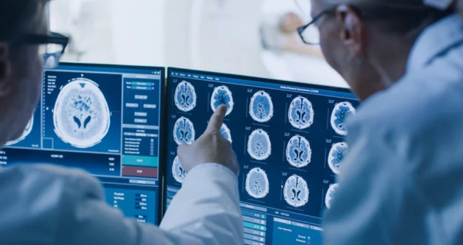
Developments like whole-body MRI and dedicated scanners within treatment environments are making imaging more accessible and effective. Meanwhile, the shift toward patient-centric care pathways reflects a broader goal of combining technological innovation with practical solutions to improve diagnostic workflows in medicine.
MRI in Oncology: Emerging Applications
MRI technology continues to evolve, playing a critical role in cancer detection, treatment planning, and monitoring. Improvements in imaging quality and novel MRI techniques have expanded its capability, providing detailed tumor characterization and enabling functional and molecular insights. These advancements support more tailored approaches in oncology care.
Advances in Tumor Detection and Characterization
MRI provides superior soft tissue contrast, making it highly effective for detecting tumors in organs like the brain, prostate, and gastrointestinal tract. Recent developments include higher-field MR scanners (1.5 T to 3 T), which improve spatial resolution and image clarity.
Whole-body MRI (WB-MRI) is increasingly used for early diagnosis and comprehensive cancer staging, with sequences such as Dixon and diffusion-weighted imaging (DWI) enhancing lesion detection. This method avoids ionizing radiation exposure, offering a safer alternative for repeated assessments.
MRI’s ability to differentiate tissue types based on proton density, T1, and T2 characteristics aids in precise tumor delineation. This is crucial for identifying tumor boundaries and heterogeneity, guiding surgical and radiation therapy decisions effectively.
Functional and Molecular Imaging
Functional MRI techniques, including diffusion-weighted imaging and dynamic contrast-enhanced MRI, provide information beyond anatomy by assessing tumor cellularity, vascularity, and permeability. These parameters help monitor tumor aggressiveness and treatment response.
Molecular imaging with MRI is gaining importance, combining traditional structural imaging with biomarkers that track molecular and metabolic changes in tumors. This enables detection of therapy targets and early identification of resistance mechanisms.
Hybrid modalities like PET/MRI merge metabolic data from PET with high-resolution MRI images, offering comprehensive insight into tumor biology and heterogeneity. This integration supports better treatment planning and response evaluation.
Personalized Oncology Using MRI Data
MRI data contributes significantly to personalized cancer treatment by refining tumor characterization and monitoring therapeutic effects in real time. Radiotherapy planning benefits from MRI’s precise delineation of tumors and surrounding healthy tissues, reducing collateral damage.
Advanced MRI techniques allow prediction of treatment response, enabling oncologists to tailor therapies based on individual tumor biology. Imaging biomarkers derived from functional and molecular scans enhance patient stratification in clinical protocols.
Emerging MRI-based theranostics aims to integrate diagnosis and therapy, using MRI-visible agents to guide and assess targeted treatments. This approach holds promise for more effective, individualized cancer management.
MRI in Cardiology: Innovations and Developments
Cardiac MRI continues to advance with enhanced imaging techniques, faster acquisition times, and deeper integration of artificial intelligence. These developments enable detailed tissue analysis, improved risk assessment, and precise evaluation of vascular conditions.
Myocardial Tissue Characterization
Innovations in myocardial tissue characterization focus on improving the accuracy and speed of detecting fibrosis, edema, and inflammation. Techniques like T1 and T2 mapping provide quantitative measures, enabling clinicians to distinguish between healthy and diseased myocardial tissue with greater precision.
AI-driven reconstruction methods reduce noise and artifacts, improving image clarity. These advances help in diagnosing cardiomyopathies and myocarditis non-invasively. Moreover, native T1 mapping without contrast agents is increasingly used, benefiting patients with renal impairments.
Cardiac MRI for Risk Stratification
Cardiac MRI aids in stratifying risk by providing detailed insights into cardiac function and tissue viability. Late gadolinium enhancement (LGE) remains essential for detecting scar tissue and predicting arrhythmia risk.
New AI algorithms analyze large datasets combining MRI, genetic, and clinical data to personalize risk profiles. This approach supports decision-making in heart failure management and guides interventions by identifying patients at higher risk for adverse events.
Assessment of Vascular Disease
MRI techniques now allow comprehensive evaluation of vascular diseases such as atherosclerosis and aneurysms without ionizing radiation. Advanced flow imaging measures wall shear stress and detects plaque vulnerability.
3D imaging sequences provide high-resolution views of coronary arteries and large vessels. AI assists in motion artifact correction and automated segmentation, enhancing diagnostic accuracy and monitoring vascular remodeling over time.
MRI in Neurology: Novel Techniques and Clinical Impact
Advancements in MRI technology have enhanced the precision and scope of neurological imaging. These developments improve the detection and monitoring of brain structure, degeneration, and cognitive function, offering critical insights for diagnosis and treatment.
High-Resolution Brain Imaging
High-resolution MRI enables detailed visualization of brain anatomy, enhancing the detection of subtle structural abnormalities. This technique leverages stronger magnetic fields and improved coil designs to produce images with finer spatial resolution.
It is particularly effective for identifying small lesions, microbleeds, and early changes in brain tissue that were previously undetectable. High-resolution imaging supports surgical planning and disease monitoring by precisely mapping affected areas. The increase in image clarity aids clinicians in diagnosing complex neurological conditions with greater confidence.
Neurodegenerative Disease Assessment
Advanced MRI methods provide quantitative measures of brain tissue changes associated with neurodegenerative diseases such as Alzheimer’s, Parkinson’s, and multiple sclerosis.
Techniques like volumetric analysis, diffusion tensor imaging (DTI), and iron mapping quantify atrophy, white matter integrity, and abnormal mineral deposits. These biomarkers enable earlier detection compared to traditional methods and improve disease staging and progression tracking. Clinicians can also better evaluate treatment efficacy over time.
This quantitative approach assists in differentiating between overlapping clinical syndromes and tailoring personalized care strategies based on the extent and pattern of neurodegeneration.
Functional MRI for Cognitive Disorders
Functional MRI (fMRI) measures brain activity by detecting blood flow changes, providing insights into neural network function and cognitive processes.
It plays a growing role in understanding disorders such as dementia, epilepsy, and traumatic brain injury by mapping functional impairments and compensatory mechanisms. fMRI helps guide interventions by identifying regions critical for language, memory, and executive functions.
Current research focuses on improving temporal and spatial resolution to capture more precise neural dynamics. This aids in both diagnosis and evaluating responses to therapeutic interventions targeting cognitive functions.
AI and Machine Learning in MRI
Artificial intelligence and machine learning are transforming MRI by improving image interpretation, enhancing diagnostic accuracy, and streamlining clinical workflows. These technologies apply complex algorithms to extract valuable insights from MRI data, supporting decision-making in oncology, cardiology, and neurology.
Automated Image Analysis
AI systems enable automated analysis of MRI scans, identifying abnormalities such as tumors, plaques, or lesions with high precision. Deep learning models process raw imaging data to segment tissues, detect pathologies, and quantify structural changes, reducing the need for manual intervention.
In oncology, automated image analysis aids in tumor characterization and volumetric assessment, improving treatment planning. In cardiology, it facilitates accurate mapping of heart function and scar tissue. Neurological applications include lesion detection in multiple sclerosis and brain atrophy measurement in neurodegenerative diseases.
These tools reduce human error and analysis time, allowing radiologists to focus on complex cases. They also enhance reproducibility across institutions by standardizing image interpretation.
Predictive Modeling for Disease Outcomes
Machine learning predicts disease progression by analyzing patterns in MRI data combined with clinical variables. Models forecast outcomes such as tumor growth, cardiac events, or cognitive decline based on imaging biomarkers.
In oncology, AI-driven modeling enables personalized prognosis and treatment response prediction. Cardiac MRI data integrated with AI can stratify patients at risk for heart failure or arrhythmias. In neurology, predictive algorithms assess the likelihood of disease exacerbation or response to therapies.
These models support earlier interventions by identifying high-risk patients. Continuous refinement with large datasets enhances predictive accuracy over time, contributing to precision medicine.
Workflow Optimization and Efficiency
AI optimizes MRI workflows by accelerating image acquisition and automating repetitive tasks. Techniques like compressed sensing combined with deep learning reduce scan times without compromising image quality, improving patient comfort and throughput.
Automated image reconstruction and quality control streamline data processing, allowing faster report generation. Scheduling and protocol selection can be enhanced by AI to tailor scans to individual patient needs.
This efficiency lowers operational costs and increases scanner availability. It also reduces bottlenecks in busy clinical settings, fostering timely diagnosis and treatment across oncology, cardiology, and neurology.
Technological Advances in MRI Hardware and Software
MRI technology continues to evolve through improvements in magnet strength, device portability, and image processing techniques. These developments focus on enhancing diagnostic precision, expanding access to imaging, and reducing risks associated with scans.
Ultra-High Field MRI Systems
Ultra-high field MRI systems, typically operating at 7 Tesla (T) or above, provide significantly higher signal-to-noise ratios compared to standard 1.5T or 3T machines. This increase enables more detailed visualization of soft tissues, benefiting oncology, cardiology, and neurology diagnosis.
The enhanced spatial and contrast resolution improves the detection of small lesions and subtle structural abnormalities. Researchers are also exploring ultra-high field MRI for functional imaging and spectroscopy, providing insights into metabolic processes.
Challenges include increased susceptibility artifacts and higher operational costs. However, ongoing hardware optimization and software corrections aim to mitigate these issues. Overall, ultra-high field MRI represents a critical advancement for complex clinical and research applications.
Portable and Point-of-Care MRI Devices
Portable MRI devices have emerged to provide imaging in non-traditional settings, including rural areas and bedside use. These systems usually operate at lower magnetic fields (typically below 1T), trading off some image resolution for mobility and accessibility.
Portable MRIs allow rapid assessment without requiring patient transport to specialized imaging centers, which is especially valuable in emergency care and intensive care units. New developments include lightweight magnets and streamlined software interfaces designed for quick operation.
While image quality does not yet match that of higher-field systems, continuous improvements in coil design and reconstruction algorithms enhance clinical utility. This technology expands the reach of MRI diagnostics to more patients and diverse healthcare environments.
Dose Reduction and Image Quality Improvements
MRI inherently avoids ionizing radiation, which is critical in imaging-sensitive populations. Nevertheless, optimizing scan protocols to reduce acquisition time and increase image clarity remains a priority.
Advanced software algorithms, including AI-based reconstruction methods, enable faster image acquisition with maintained or improved quality. Techniques such as compressed sensing and parallel imaging reduce scan times and motion artifacts.
Hardware improvements, like more efficient gradient coils and improved RF coil arrays, contribute to sharper images and reduced noise. Together, these advancements enhance patient comfort and throughput without compromising diagnostic accuracy.
Interdisciplinary Collaboration and Integration
Advancements in MRI technology require seamless integration of multiple imaging methods and close cooperation between radiology and clinical teams. This collaboration enhances diagnostic accuracy and patient care across oncology, cardiology, and neurology.
Multi-Modality Imaging Approaches
Multi-modality MRI combines various imaging sequences to provide a detailed and comprehensive view of tissue structures and function. This approach is crucial in oncology, cardiology, and neurology for detecting subtle abnormalities that single imaging techniques might miss.
By merging anatomical, functional, and molecular data, multi-modality imaging improves diagnosis and monitoring of disease progression. For example, in oncology, integrating diffusion-weighted imaging (DWI) with contrast-enhanced MRI helps differentiate tumor types and evaluate treatment response.
The use of artificial intelligence to fuse and analyze these diverse datasets is growing, enabling more precise interpretation and minimizing human error. This comprehensive imaging strategy supports personalized treatment planning and better patient outcomes.
Collaboration Between Radiology and Clinical Teams
Effective collaboration between radiologists and clinical teams is essential for translating MRI data into actionable clinical decisions. Radiologists provide expertise on imaging interpretation, while clinicians contribute patient history and treatment context.
Regular interdisciplinary meetings foster real-time communication, ensuring that imaging findings align with clinical objectives. This is especially important in cardio-oncology, where cardiovascular risks intersect with cancer treatments.
Training programs that include clinicians, radiologists, and allied health professionals promote shared understanding and improve workflow. Such collaboration bridges knowledge gaps, facilitating timely diagnosis and integrated patient management.
Challenges and Future Perspectives in MRI Research
Magnetic resonance imaging continues to evolve across oncology, cardiology, and neurology, yet several key hurdles remain. These include the need for uniform data standards and navigating complex ethical issues around patient data use. Addressing these challenges is crucial to advancing MRI research and clinical impact.
Standardization and Regulatory Considerations
MRI research faces significant variability due to different scanner technologies, imaging protocols, and data processing methods. This inconsistency complicates multi-center studies and the integration of MRI-derived biomarkers in clinical practice.
Regulatory bodies require clear validation and reproducibility standards before approving MRI-based tools for diagnosis or treatment monitoring. Harmonizing imaging protocols and establishing universal quality control measures will improve data comparability and facilitate regulatory acceptance.
Efforts toward standardized reporting frameworks and collaborative networks aim to align practices globally. Such measures are essential for translating advanced MRI methods—like multimodal imaging and AI-driven metrics—into routine patient care.
Ethical and Privacy Issues in MRI Data Usage
MRI datasets often contain sensitive patient information, requiring strict safeguards to protect privacy. With increasing use of artificial intelligence, large-scale data sharing and secondary data use raise concerns about consent, anonymization, and potential misuse.
Ethical frameworks must ensure transparency in data handling and secure informed consent processes for future research applications.
Institutions adopt robust data governance policies emphasizing encryption, access control, and ongoing oversight. These practices balance innovation needs with patient rights, enabling responsible MRI research while mitigating privacy risks.
Also Read :