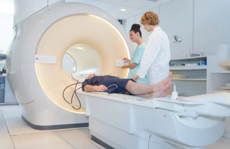
Cardiovascular disease remains the leading cause of death worldwide, driving an urgent demand for technologies that enable earlier detection, accurate diagnosis, and personalized treatment. Among the tools reshaping cardiology, Cardiac Magnetic Resonance Imaging (Cardiac MRI or CMR) stands at the forefront.
Once limited to research centers, Cardiac MRI has evolved into a mainstream clinical powerhouse, capable of providing comprehensive insights into the heart’s anatomy, function, and tissue health—without radiation exposure or invasive procedures. As artificial intelligence (AI), machine learning, and molecular imaging merge with MRI technology, we are entering a new era where Cardiac MRI will define the future of cardiovascular medicine.
This article explores how Cardiac MRI is transforming diagnosis, treatment planning, and patient outcomes—while shaping the next generation of precision cardiology.
The Power of Cardiac MRI: A Complete View of the Heart
Cardiac MRI is unique in its ability to capture high-resolution, three-dimensional images of the heart in motion. Unlike CT scans or echocardiography, it provides unparalleled detail about both structure and tissue composition, allowing physicians to assess not just how the heart looks—but how it functions at the cellular level.
Key Advantages of Cardiac MRI Include:
- Radiation-Free Imaging: Safe for repeated follow-up examinations.
- Superior Soft-Tissue Contrast: Enables visualization of heart muscle, valves, and blood flow in exquisite detail.
- Quantitative Analysis: Provides measurable data on heart function, perfusion, and tissue composition.
- Comprehensive Assessment: Detects congenital defects, ischemia, fibrosis, inflammation, and scarring in a single session.
In the coming decade, Cardiac MRI will move beyond structural imaging to become a real-time, data-driven tool guiding every step of cardiovascular care.
Advancements Redefining Cardiac MRI Technology
Recent breakthroughs in MRI engineering and software have dramatically enhanced image quality, speed, and diagnostic capability.
1. Accelerated Scanning Techniques
New acquisition methods—such as compressed sensing and parallel imaging—reduce scan times from 45 minutes to under 10, making Cardiac MRI more accessible and patient-friendly.
2. 4D Flow MRI
This innovation allows visualization of blood flow dynamics throughout the heart and vessels, mapping patterns that reveal early signs of valve disease or vascular abnormalities long before symptoms appear.
3. T1 and T2 Mapping
Quantitative tissue mapping detects subtle changes in myocardium (heart muscle) due to inflammation, edema, or fibrosis—key indicators of heart failure or myocarditis.
4. Low-Field and Portable MRI
Emerging low-field systems are smaller, cheaper, and easier to install, expanding access to advanced cardiac imaging even in resource-limited regions.
These advances mark the transition of Cardiac MRI from a diagnostic tool to a predictive, personalized medicine platform.
Early Diagnosis: Detecting Cardiovascular Disease Before It Strikes
One of the most powerful aspects of modern Cardiac MRI is its ability to detect disease at the earliest stages, often before structural damage or clinical symptoms occur.
Applications in Early Detection Include:
- Ischemic Heart Disease: MRI perfusion imaging identifies areas of reduced blood flow with high accuracy.
- Myocarditis and Pericarditis: Tissue characterization sequences detect inflammation, guiding appropriate treatment.
- Cardiomyopathies: MRI reveals early signs of hypertrophic, dilated, or restrictive cardiomyopathy through tissue mapping.
- Atherosclerosis: Vessel wall imaging detects plaque composition and vulnerability before it leads to heart attack or stroke.
By identifying these silent precursors, MRI helps clinicians intervene proactively, preventing irreversible damage and improving long-term outcomes.
MRI in Heart Failure Management
Heart failure affects over 60 million people globally, and Cardiac MRI plays a pivotal role in understanding its causes and progression.
- Functional Analysis: MRI precisely measures ejection fraction, wall motion, and ventricular volumes.
- Tissue Viability Assessment: Late gadolinium enhancement (LGE) identifies areas of scar tissue, helping determine whether damaged myocardium can recover after revascularization.
- Fibrosis Detection: T1 mapping detects diffuse fibrosis linked to poor prognosis, guiding therapy choices.
- Therapy Monitoring: MRI evaluates response to medications, cardiac resynchronization therapy (CRT), and regenerative treatments.
In essence, Cardiac MRI provides the complete story of heart failure—from cause to cure—making it the gold standard for patient-tailored management.
MRI-Guided Cardiac Interventions: Precision in Action
The next frontier in cardiology is MRI-guided intervention—performing minimally invasive procedures with real-time MRI visualization instead of X-ray fluoroscopy.
Emerging MRI-Guided Applications Include:
- Catheter-Based Repairs: MRI visualizes catheters and guidewires inside the heart without radiation.
- Ablation Therapy: MRI monitors heat distribution during arrhythmia treatments to ensure precise targeting.
- Cardiac Biopsies: Real-time imaging ensures accurate sampling of affected tissue.
- Valve and Stent Placement: MRI provides detailed anatomical guidance during complex procedures.
These MRI-guided interventions combine surgical precision with imaging intelligence, reducing risks and improving outcomes for complex cardiovascular conditions.
Cardiac MRI in Pediatric and Congenital Heart Disease
For children and patients with congenital heart defects, minimizing radiation exposure is crucial. Cardiac MRI offers a safe, non-invasive, and highly accurate imaging option that can evaluate structural anomalies, flow patterns, and surgical repairs across all age groups.
In pediatric cardiology, MRI helps:
- Track heart growth and post-surgical outcomes.
- Assess shunts, valve malformations, and vessel anomalies.
- Monitor long-term complications without repeated radiation scans.
As scan times shorten and patient comfort improves, Cardiac MRI will become the preferred standard for congenital heart disease monitoring worldwide.
AI and Cardiac MRI: Intelligent Insights for Precision Medicine
Artificial intelligence is revolutionizing every step of the Cardiac MRI workflow—from acquisition to interpretation.
AI Innovations in Cardiac MRI Include:
- Automated Image Segmentation: AI rapidly outlines cardiac chambers and vessels, reducing reporting time.
- Predictive Modeling: Machine learning algorithms forecast risk of heart failure, arrhythmia, or sudden cardiac death.
- Personalized Diagnostics: AI links imaging biomarkers to genetic and clinical data for tailored treatment plans.
- Automated Quality Control: Ensures consistent imaging standards across hospitals.
The integration of AI transforms MRI from a diagnostic snapshot into a real-time decision support system, empowering cardiologists to deliver faster and more precise care.
The Role of MRI in Cardio-Oncology
As cancer therapies improve, cardiotoxicity—heart damage caused by chemotherapy or radiation—has become a growing concern. Cardiac MRI is emerging as the gold standard for detecting and monitoring heart damage in cancer survivors.
- Early Detection: T1/T2 mapping identifies subtle myocardial injury before functional decline.
- Long-Term Monitoring: MRI evaluates changes in cardiac performance throughout and after cancer treatment.
- Therapy Adjustment: Real-time insights help oncologists adapt chemotherapy regimens to minimize cardiac risk.
This cross-disciplinary field, known as cardio-oncology, exemplifies how MRI bridges specialties to protect heart health while advancing cancer care.
Future Applications: The Next Generation of Cardiac MRI
As technology evolves, the future of Cardiac MRI will be defined by speed, intelligence, and personalization.
Key Trends Shaping the Future:
- Ultra-Fast MRI: Whole-heart imaging in seconds, enabling cardiac screening for large populations.
- AI-Driven Predictive Imaging: Algorithms will predict cardiac events years before they occur.
- Molecular MRI: Detects cellular-level changes, offering insight into early atherosclerosis and heart failure.
- Hybrid Imaging (PET/MRI): Combines metabolic and structural data for comprehensive cardiac assessment.
- Wearable MRI Technology: Research is underway into portable, low-field MRI systems for on-demand cardiac monitoring.
These innovations promise to make cardiac MRI not just a diagnostic modality—but a core component of preventive and precision cardiology.
Challenges and the Road Ahead
Despite its enormous potential, the widespread adoption of Cardiac MRI faces a few challenges:
- High Costs and Infrastructure Needs for advanced MRI systems.
- Lengthy Scan Times in conventional setups, though AI is reducing this significantly.
- Training Requirements for radiologists and cardiologists in advanced image interpretation.
However, as costs fall and automation rises, these challenges are steadily diminishing. The future will see Cardiac MRI systems that are faster, smarter, and more widely accessible, even in community hospitals and mobile clinics.
Conclusion: The Heart of Precision Medicine
Cardiac MRI is more than a diagnostic innovation—it is the beating heart of the future of cardiovascular medicine. With its unmatched ability to visualize, quantify, and predict heart disease, it bridges the gap between imaging, data, and treatment.
As AI integration deepens and technology advances, Cardiac MRI will transform from a specialized imaging technique into an everyday clinical necessity—guiding prevention, treatment, and recovery across every stage of heart health.
In the next decade, hospitals will evolve into MRI-guided cardiovascular centers, where imaging doesn’t just reveal disease—it directs personalized therapy, improves outcomes, and saves lives.
The future of Cardiac MRI is not just about seeing the heart—it’s about understanding it like never before.
Also Read :
- How MRI Is Transforming Cancer Diagnosis and Therapy
- MRI in Neurology: Future Applications for Brain Health
- The New Standard: MRI-Guided Treatment Departments
