Magnetic Resonance Imaging (MRI) is one of the most trusted diagnostic technologies in modern healthcare. It plays a vital role in detecting, staging, and monitoring cancer, as well as evaluating a wide range of non-cancerous conditions. Despite its proven clinical value, questions about MRI and cancer safety continue to circulate among patients and the general public. Concerns often stem from misunderstandings about exposure levels, magnetic fields, and contrast agents.
This in-depth cancer safety review examines how MRI works, whether it poses any cancer risk, what large scientific studies conclude, and why MRI is widely considered a low-risk imaging modality. The goal is to provide clear, evidence-based answers in a way that is easy to understand and medically accurate.
Understanding Magnetic Resonance Imaging (MRI)
How MRI Technology Works
MRI uses a combination of strong magnetic fields, radiofrequency (RF) pulses, and advanced computer processing to generate detailed images of the body’s internal structures. Unlike X-rays or CT scans, MRI does not rely on ionizing radiation.
The process involves:
- Aligning hydrogen atoms in the body using a powerful magnetic field
- Applying RF energy to disturb this alignment
- Capturing the signals released as atoms return to their original state
These signals are converted into high-resolution images of soft tissues, organs, and blood vessels.
MRI vs. Radiation-Based Imaging
A key reason MRI is considered safe in cancer care is its lack of ionizing radiation. Ionizing radiation has enough energy to damage DNA, which can increase cancer risk over time. MRI uses non-ionizing energy, which does not alter genetic material or initiate cancer-causing cellular changes.
MRI and Cancer Risk: What Does the Science Say?
Is MRI Linked to Cancer Development?
Extensive research over several decades has found no evidence linking MRI scans to cancer development. Large population studies, long-term patient follow-ups, and occupational research involving MRI technicians all support the same conclusion: MRI does not increase cancer risk.
Key findings include:
- No association between MRI exposure and tumor formation
- No increase in cancer rates among patients undergoing repeated MRIs
- No genetic or DNA damage linked to MRI magnetic fields
As a result, MRI is often preferred for patients who require frequent imaging, including cancer survivors.
Why MRI Is Used in Cancer Diagnosis and Monitoring
MRI is widely used in oncology because it offers:
- Exceptional soft tissue contrast
- Precise tumor localization
- Accurate assessment of tumor size and spread
- Safe long-term monitoring without radiation exposure
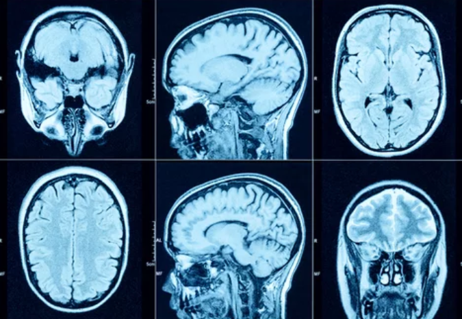
For cancers of the brain, spine, liver, prostate, and female reproductive organs, MRI is often the imaging method of choice.
MRI Exposure Levels and Safety Standards
Magnetic Field Strength and Regulation
Clinical MRI scanners typically operate at:
- 1.5 Tesla (standard clinical use)
- 3 Tesla (higher resolution imaging)
These magnetic field strengths are strictly regulated by international safety organizations. Research-grade scanners with higher field strengths are used only under controlled conditions and with ethical approval.
Radiofrequency Energy and Tissue Heating
RF energy used during MRI can cause minimal tissue heating, measured by the Specific Absorption Rate (SAR). Modern MRI systems continuously monitor SAR levels to ensure patient safety.
Important points include:
- SAR limits are set far below harmful thresholds
- Automatic controls prevent overheating
- Patients rarely experience more than mild warmth
There is no evidence linking RF exposure during MRI to cancer or long-term health effects.
MRI Contrast Agents and Cancer Safety
Understanding MRI Contrast Media
Some MRI scans use contrast agents, most commonly gadolinium-based contrast agents (GBCAs), to enhance image clarity. These agents help distinguish tumors, inflammation, and abnormal blood vessels.
Gadolinium and Cancer Concerns
Research has shown that gadolinium contrast agents:
- Do not cause cancer
- Are rapidly eliminated in patients with normal kidney function
- Have a strong safety record when used appropriately
Although trace gadolinium retention has been detected in some tissues, no clinical harm or cancer-related outcomes have been linked to this finding. Ongoing research continues to improve contrast agent formulations and safety guidelines.
MRI Contrast Use in Cancer Patients
For cancer patients, contrast-enhanced MRI is often essential for:
- Accurate tumor staging
- Assessing treatment response
- Detecting recurrence
Physicians carefully evaluate the benefits and risks before administering contrast, especially in patients with kidney disease.
MRI in Children and Cancer Risk
Pediatric MRI Safety
MRI is commonly used in children due to its radiation-free nature. Pediatric imaging guidelines frequently recommend MRI over CT scans whenever possible.
Long-term studies show:
- No increased cancer risk in children exposed to MRI
- No adverse developmental or neurological outcomes
- Excellent safety for repeated imaging
This makes MRI particularly valuable for children with chronic illnesses or cancer requiring ongoing monitoring.
MRI During Pregnancy and Cancer Safety
Is MRI Safe for Pregnant Patients?
MRI is generally considered safe during pregnancy when medically necessary. Research has found:
- No evidence of fetal harm
- No increased cancer risk later in life
- No DNA damage associated with prenatal MRI exposure
Contrast agents are typically avoided unless the diagnostic benefit clearly outweighs potential risks.
High-Field MRI and Cancer Risk
Does Higher Tesla Mean Higher Risk?
Higher-field MRI scanners, such as 3T systems, provide improved image quality but remain within established safety limits. Scientific evidence shows:
- No increase in cancer risk compared to standard MRI
- Controlled RF exposure
- No long-term adverse health outcomes
High-field MRI is widely used in advanced cancer imaging and research.
Comparing MRI With Other Imaging Techniques
MRI vs. CT Scans in Cancer Care
| Feature | MRI | CT Scan |
|---|---|---|
| Ionizing radiation | No | Yes |
| Cancer risk | None | Low but measurable |
| Soft tissue detail | Excellent | Moderate |
| Repeat imaging safety | Very high | Dose-dependent |
This comparison highlights why MRI is often preferred for long-term cancer surveillance.
Common Myths About MRI and Cancer
Myth 1: MRI Can Cause Cancer
Fact: MRI does not use radiation and has not been linked to cancer.
Myth 2: Repeated MRI Scans Are Dangerous
Fact: There is no cumulative exposure risk from MRI scans.
Myth 3: MRI Alters DNA
Fact: Magnetic fields and RF waves do not damage genetic material.
Psychological and Physical Effects of MRI
Short-Term Sensations
Some patients may experience:
- Loud noises during scanning
- Mild warmth
- Temporary discomfort from remaining still
These effects are short-lived and unrelated to cancer or long-term health risks.
Anxiety and Claustrophobia
Emotional discomfort is common but manageable through:
- Open MRI systems
- Shorter scan times
- Relaxation techniques and patient education
These measures improve patient experience without affecting safety.
Why MRI Is a Cornerstone of Cancer Care
MRI has become essential in oncology because it combines:
- High diagnostic accuracy
- Exceptional safety
- Non-invasive imaging
- Long-term reliability
Its ability to detect cancer early and monitor treatment without exposing patients to radiation makes it a cornerstone of modern cancer care.
Final Verdict: MRI and Cancer Safety
After decades of research and clinical use, the conclusion is clear: Magnetic Resonance Imaging does not increase cancer risk. MRI’s non-ionizing technology, strict safety standards, and extensive scientific validation make it one of the safest diagnostic imaging tools available.
For patients concerned about cancer safety, MRI offers reassurance, precision, and peace of mind. Whether used for diagnosis, treatment planning, or long-term monitoring, MRI continues to deliver life-saving insights without compromising long-term health.
Also Read :