Neuroimaging and neurosurgical approaches in stroke recovery continue to be advanced in treatment and rehabilitation. Recovery from a stroke is quite complex, with many medical techniques and approaches adopted for it. The neuroimaging and neurosurgical methods have, undoubtedly, made a great contribution to helping the patients regain their lost functions of the body. The procedures provide insight into the brain functions and allow focused interventions that may significantly enhance outcomes.
Understanding the response of the brain after stroke is important. Neuroimagings help the doctors visualize changes in the brain and pinpoint the most affected areas. This is importantly needed when planning effective neurosurgical procedures that may help in rehabilitation.
The combination of neuroimaging with surgical technique opens a new horizon in stroke recovery. Thus, individualized treatment plans are carried out to meet the patient’s needs and make a gain toward independence and a better quality of life.
Key Takeaways
- The neuroimaging technique helps to show changes that occur within the brain after a stroke.
- Neurosurgical interventions may be provided to aim at the specific site within the brain to bring about better stroke recovery.
- Treatment approaches need to be tailored to each individual in stroke rehabilitation.
Fundamentals of Stroke and Neuroimaging
Stroke is a severe pathological condition that has the potential to cause devastating disability. Neuroimaging plays a very important role in understanding stroke and guiding appropriate treatment options. Several imaging modalities are helpful for a doctor to visualize changes in the brain that would help in effective management for recovery from stroke.
Stroke Type and Pathophysiology
There are primarily two kinds of stroke: ischemic and hemorrhagic. An ischemic stroke involves blockage of a blood vessel supplying the brain. This is typically due to a blood clot, in which case the oxygen supply to a particular section of the brain is inadequate and may consequently be damaged.
Hemorrhagic strokes are those that occur due to leakage or rupture of the blood vessel in the brain, resulting in bleeding in or around the brain. It can be due to high blood pressure and aneurysms. Knowledge of the type of stroke will help in understanding the necessary mode of treatment.
Both the types of stroke present different symptoms and brain injuries, which influence various recovery and rehabilitation methods accordingly.
Neuroimaging for Stroke Diagnosis: How It Has Evolved
Neuroimaging technology has rapidly evolved during the last years. New methods allow faster and more specific diagnostics of strokes. These improvements involve the enhancement of MRI and CT scans to pick up subtle changes in the brain tissue appearing little time after stroke onset.
Recent technologies like Diffusion Weighted Imaging detect ischemic strokes in a matter of minutes, which is very important for its effective treatment since the sooner the intervention, the better the outcome. Also, CTA, or CT Angiography visualizes the blood vessels and helps in making decisions whether to perform surgery to remove clots or repair vessels.
Neuroimaging Modalities
There are several neuroimaging modalities that are used in stroke evaluation.
- CT Scans: This is usually the first imaging test done to rapidly identify hemorrhagic strokes.
- MRI: It gives suitable structural pictures of the brain tissue. This modality is beneficial, especially for the assessment of early ischemic changes.
- Ultrasound: It is used in the assessment of blood flow via carotid arteries to assist in the estimation of stroke risk.
All these techniques have some strengths and weaknesses. For instance, though CT is fast and also more available, yet the details of MRI are finer and broader. Knowledge of these techniques definitely assists health professionals in the management and care of patients for better recovery post-stroke.
Neurosurgical Interventions in Stroke Recovery
Approaches to neurosurgery also play a significant role in the improvement of recovery after stroke. Techniques include the use of minimally invasive procedures, rehabilitative neurosurgery, and advanced brain-computer interfaces which contribute well in the quality of life of patients.
Minimally Invasive Techniques
These minimally invasive techniques minimize the time a person has to recover and therefore result in better outcomes for the stroke patient. Small incisions are performed by the surgeons and guides enabled with advanced imaging to accurately place the instruments at the site. This, in turn, causes less tissue damage, and with that, the complications associated are hugely reduced.
Some general forms of minimally invasive techniques include endovascular thrombectomy, which removes blood clots from the brain. Several hospitals are reporting that patients treated through these techniques have shorter stays and quicker rehabilitation times.
Besides this, these surgeries can often be carried out under local anesthesia. In such types of anesthesia, the patient remains awake and is thus able to provide immediate feedback during the surgery.
Rehabilitative Neurosurgery
Rehabilitative neurosurgery aims at improving recovery by specific interventions. This may include the placement of neuromodulation devices or specific surgical adjustments in the brain pathways to aid in recovery.
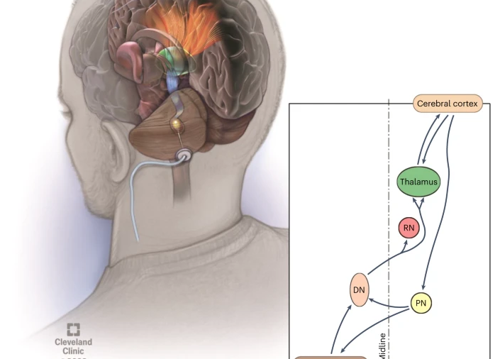
One such example is deep brain stimulation. With the use of implanted electrodes in selected areas of the brain, DBS can help stroke patients in their motor improvement. This modality allows patients to have further mobility with increased coordination so as to continue with everyday activities.
Studies show that rehabilitative neurosurgery, when applied in conjunction with conventional therapy, can have a better outcome. Synergy between surgical and conservative approaches improves advances, which is often crucial in rehabilitation.
Brain-Computer Interface in Rehabilitation
BCIs represent an innovative tool in stroke recovery. These systems connect the brain directly to external devices, which can allow patients to control computers or prosthetics with their thoughts.
BCIs can help the patient practice and get feedback in regaining motor functions; for instance, they can be used to train movements in a paralyzed limb. While patients imagine moving, the device translates those actions into performance that helps develop neural pathways.
Clinical trail results are promising, as both the motor skills and the quality of life improve. For many, BCI does provide a hint of hope toward rehabilitation.

Also Read :
