
Functional MRI in Cognitive Neurosurgery: Development and Uses
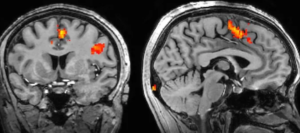
The new modality of neuroimaging called fMRI has proven itself revolutionary in cognitive neurosciences. Its unique advantage of viewing brain activity instantly has enhanced its utility in surgical planning and has improved the outcomes for patients. By offering a diagram of the active parts of the brain, fMRI gives critical information that can lead to making decisions during complicated cognitive surgery.
This integration helps in identifying critical functional areas and minimizing damage to important brain tissues in cognitive neurosurgery. In this light, the imaging techniques provide a basis on which surgeons apply while approaching and ensuring that there is no damage affecting the cognitive functions of the brain while addressing pathological conditions. With the advancement in technology, fMRI has tended to increase in precision and utility, and thus this makes the modality important in contemporary surgical practice.
Basics and applications of fMRI show the modality’s importance in the development of cognitive neurosurgery. As this area continues to progress, appreciation of how to exploit fMRI will be increasingly germane both to the clinician and the researcher.
Key Points
- fMRI is the main method of visualizing brain function for surgical planning.
- It provides the identification of eloquent areas of the brain that should be spared during surgical interventions.
- Ongoing developments increase its usefulness in cognitive neurosurgery.
Principles of Functional MR
fMRI has become an important tool in the cognitive neurosciences and thousands of studies have used the technique to examine the neural correlates of cognitive processes. Technology detects changes in blood flow in order to find areas of the brain that are more active during particular tasks, thus offering a view into both cognitive processes and brain organization.
Principles of fMRI Imaging
The basic principle of fMRI rests on the so-called Blood Oxygen Level Dependent (BOLD) contrast mechanism: during brain activation, the metabolic demand for oxygen increases; as a consequence, more blood flows to the activated area. This, in turn, causes changes in the relative proportion of oxyhemoglobin and deoxyhemoglobin, and this is what fMRI detects.
The images are generated by measuring signal variations produced by changing amounts of oxygenated blood. It has the advantage of high temporal and spatial resolution mapping of brain activity. fMRI allows observation of very rapid changes in neural activity and thus can be used to see the brain while it works on cognitive tasks.
fMRI vs. Traditional Neuroimaging
While traditional neuroimaging methods, such as CT or MRI, are capable of providing structural pictures, fMRI provides dynamic data on the functioning aspects of the brain. Other techniques, such as positron emission tomography, require radioactive tracers and result in slower temporal resolution.
fMRI represents a noninvasive technique with no ionizing radiation, hence safer for repeated measures. This is because it uniquely allows for the mapping of brain activity in various conditions, thus extending the ability to study cognitive processes beyond static imaging techniques.
Role in the Understanding of Brain Function
fMRI has greatly increased research into brain functionalities such as memory, attention, and perception. Research using fMRI has been able to show how different brain networks cooperate during complicated tasks.
It helps in the design of focused interventions for neurological disorders by the identification of regions involved in specific functions. Secondly, it provides various tools that assess techniques/therapies aimed at enhancing cognition, hence bridging neuroscience and clinical applications.
Application in Cognitive Neurosurgery
fMRI has become an integral part of surgical planning for surgeries performed in cognitive neurosurgery, thus enhancing precision of surgical procedures. Its application spans preoperative planning to intraoperative decision making, including postoperative assessment to help a comprehensive approach to patient care.
Pre-Surgical Planning
The use of fMRI in presurgical planning maps brain activity regarding important functions such as language and motor. This imaging technique helps surgeons avoid eloquent areas of the brain during tumor resection or epilepsy surgery.
It maps out areas of the brain responsible for speech and comprehension, while making sure those areas are spared. This helps in planning the approach to tumors near motor pathways so as to minimize postoperative deficits. Such information allows tailored surgical strategy that addresses not only the removal of pathological tissue but the preservation of essential brain functions as well.
Intraoperative Decision Making
fMRI can help during surgery in real-time decision making. Intraoperative imaging modalities are available to the surgeon in the assessment of the dynamic functional landscape of the brain.
- Neuro-navigation: Fully advanced technologies make possible the mapping of functional areas throughout the time of the procedure.
- Immediate Feedback: The surgeon will immediately receive information regarding the activity of the brain, thus being able to adjust their approach if necessary.
- This capability enhances the safety and efficacy of surgical interventions, as this representation provides a better understanding of the operative field.
Assessment and Rehabilitation after Surgery
FMRI after surgery is quite crucial in that it shows the outcome of the surgical procedure. When pre-and post-operative brain activities are compared, it helps the clinician to identify specific changes in functions.
- Recovery Assessment: FMRI can monitor reorganization at this critical time in the life cycle of brain functions so important for understanding patient recovery trajectories.
- Rehabilitation Planning: The insights gained thereby allow targeted rehabilitation strategies to meet the unique needs of the patient.
- This approach supports not only recovery but also future therapeutic interventions in the best interest of patients for their long-term management.
Also Read :