
Using MRI for Assessment of Neurosurgical Interventions for Chronic Pain: A Comprehensive Review of Techniques and Outcomes
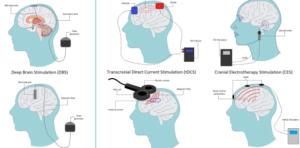
Millions of people are said to be affected by chronic pain, which most of the time is resistant to treatment. Neurosurgery offers various techniques to alleviate this pain; important in such treatments is the role played by technology in assessing these techniques. For the assessment of neurosurgical interventions in chronic pain, MRI has become a very strong tool due to its ability to render detailed images that can assist doctors in better decision-making.
Understanding MRI in this context provides a window into the brain and spine to illuminate conditions of pain. It also allows for appropriate preoperative and postoperative assessments to enable the best treatment for the patient. The use of MRI in this fashion will continue to enhance the effort to individualize interventions to ensure improved outcomes.
By researching the connection between MRI and chronic pain management, new avenues of hope can be discovered for the sufferer. Recent research demonstrates how this technology can be utilized to refine surgical procedures and, therefore, is a subject worth exploring for both medical experts and patients.
Key Points
- MRI provides important imagery that plays a vital role in assessing neurosurgical interventions.
- It helps in understanding chronic pain in a more detailed manner by illustrating its source.
- Such tailored treatments can result in better pain management outcomes.
Principles of MRI Technology in Neurosurgery
Magnetic Resonance Imaging (MRI) is a modality of importance in neurosurgery. It provides great assistance for the physician in the visualization of the brain and spinal cord, with detail that helps diagnose and plan interventions for chronic pain. The following sections outline the physics of MRI, safety considerations, and recent developments in the technology.
Physics of Magnetic Resonance Imaging
MRI works by using strong magnetic fields and radio waves to take pictures of the inside of one’s body. In general, the underlying principle is based on hydrogen atoms of water content within the body. These hydrogen atoms, while placed under a magnetic field, align in the same field.
Then, radiofrequency pulses are passed through the patient. The energy in these pulses causes the atoms to emit signals as they return to their prior state. These signals are detected by a computer and converted into detailed images.
The advantage of MRI is that it can make high-resolution images of soft tissues. It can show the difference between normal and abnormal tissues. Therefore, it is very useful for neurosurgeons. MRI does not use ionized radiation; thus, it is a great advantage compared with other techniques such as a CT scan.
Safety and Contraindications
In MRI, a critical factor in safety is used. The very strong magnetic field may be dangerous for people with specific implants such as a pacemaker or metal fragments. A patient should indicate his medical history and implants to the doctor before starting the process of MRI.
There are general contraindications for patients to undergo an MRI. These include, but are not limited to, implants, being claustrophobic, and pregnancy in some instances. Many medical professionals show precaution and alternatives provided the situation calls for it.
Pregnant patients have to be critically evaluated to avoid any possible harm to the embryo. Similarly, personnel should make sure that no metallic items are within the MRI testing area to avoid accidents.
Recent Developments in MRI Technology
The advances in MRI are immense with the passage of time. The newer techniques include fMRI, which enables doctors to view activity in the brain while it happens. This enables them to know how the brain works when doing a certain task.
High-field MRI systems generate sharper images and faster scans. This development and technology will ensure that diagnostics become more appropriate, accurate, and patient-friendly.
Further, new image analysis software tools will further facilitate the detection and characterization of brain disorders. The future for MRI in neurosurgery, therefore, looks bright and promising, with further research freeing treatments and outcomes.
Also Read :
- MRI-Based Radiomics in Neurosurgical Outcome Prediction
- MRI Innovations in Neurosurgical Robotics and Navigation Systems
- The Role of MRI in Cranial Nerve Disorders and Neurosurgical Treatment
- Intraoperative MRI in Complex Neurosurgical Cases
- Functional MRI for Neurosurgical Planning in Brain Tumor Surgery