The Wonder of MRI: Basic Principles, Techniques, and Clinical Applications of Magnetic Resonance Imaging
Magnetic resonance imaging, better known as MRI, is one of the powerful weapons of modern medicine. Utilizing strong magnets combined with the use of radio waves, it delivers sharp details of the body’s interior. Non-invasive in nature, this technology helps doctors diagnose and monitor a number of health ailments without having to use harmful radiation.
Understanding how MRI works increases appreciation for what the modality does to help in patient healthcare. MRI diagnoses tumors, evaluates brain injuries, and is crucial in the treatment of patients. Technology coupled with medical knowledge allows MRI to be used in so many diagnostic applications.
Key Takeaways
- MRI relies on magnets and radio waves to produce images of the body.
- It is the basis of diagnosis in a wide range of medical conditions.
- Research continually improves the safety and effectiveness of MRI .
Principles of MRI Technology
MRI produces highly-detailed pictures of the body by using the principles of magnetic resonance. The major components are magnetic resonance principles, superconducting magnets and radiofrequency RF waves .
Magnetic Resonance Principles
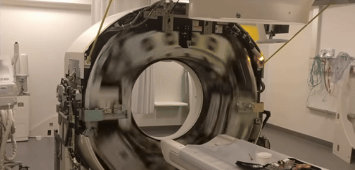
MRI technology applies the principles of nuclear magnetic resonance. It is the phenomenon where alignment of atomic nuclei, especially hydrogen, occurs in a magnetic field. When a person is taken into the tunnel of an MRI machine, hydrogen atoms in his body immediately align with the magnetic field.
Then, a pulse of RF energy is applied. This pulse will momentarily perturb the alignment of hydrogen nuclei in the body. The moment the pulse is turned off, the nuclei return to alignment and emit energy back to the scanner. This energy gets picked up by the machine and converts into images. These give high-resolution pictures of the soft tissues, which become very important for medical diagnosis.
The Role of Superconducting Magnets
MRI devices require superconducting magnets for operating, which can provide high-intensity and stable magnetic fields necessary for good quality imaging. These types of magnets involve the usage of materials at very low temperatures, which have zero electric resistance.
These machines commonly operate at temperatures of around -269 degrees Celsius. Through the process of cooling, currents can pass with no energy loss, thus producing a magnetic field several thousand times stronger than the magnetic field of Earth. The clearer the images, the stronger the magnetic field.
This feature allows the radiologist to visualize even the tiniest lesion or abnormality in the body.
RF Waves and their Function
MRI cannot take place without the use of RF waves. Once the hydrogen nuclei have aligned because of the presence of a magnetic field, then the RF coils, which generate pulsed energy, are activated. It is this energy that makes it possible to work with the hydrogen atoms.
These RF waves will cause the aligned atoms to absorb energy and jump to a higher energy state. When the RF pulse stops, the nuclei return to their prior state. As they relax, they emit energy, which the MRI machine detects.
Details collected this way help in drawing valuable pictures. Since RF works at incredible speeds and provides accurate images, MRI is one of the most favored diagnosing tools for physicians.
Clinical Applications and Safety
Medical Imaging – Magnetic Resonance Imaging: This is an important tool in health care. It is indispensable for diagnosis, and there are security features to prevent harm to the patient.
Medical Imaging in Diagnosis
MRI has many diagnostic applications in the field of medicine. It exhibits clear outlines of soft tissues, organs, and the brain. Such clarity assists the physician in locating tumors, disorders of the brain, and injuries to the joints.
Many specialists depend on MRI for accurate diagnosis. In fact, the high-resolution images can reflect changes in tissue that may not be visible with the use of any other modality. Therefore, MRI is highly valuable in neurology, orthopedics, and oncology.
Contrast Agents and Image Clarity
Sometimes, contrast agents are special substances used by doctors in order to make MRI images clearer. Such contrast agents outline certain areas within the body that might show abnormalities that would go undetected otherwise. Among the most common are gadolinium-based contrast agents.
While these agents improve the quality of the image produced, there are numerous risks with their use. Some agents can induce allergic reactions or cause kidney problems. Due to this effect, the medical history of a patient is scrutinized by doctors prior to the administration of these agents.
Safety Procedures and Protocols
The MRI is usually a safe treatment; however, some safety measures are taken to ensure patient safety. Such as the removal of metal objects from the body, since metal interferes with the magnetic field.
Moreover, technicians follow strict guidelines and regulations to avoid any kind of risk. For example, they monitor patients who may have implants of a medical nature and offer adequate shielding. Women who are pregnant should discuss their MRI safety with their providers.
Safety checks and communication between patients and their healthcare teams is also important in the managing of concerns regarding the process of a MRI for an assuring experience.

Also Read :
