The fusion of MRI and visual technology marks a significant step forward in medical imaging. By combining detailed magnetic resonance imaging with advanced visual tools, this approach creates clearer, more informative images. This fusion enhances the ability to detect and understand medical conditions by providing richer, real-time visual information.
This new era in imaging does not just improve clarity but also allows for better integration of multiple imaging methods. The combined images help doctors see conditions from different angles and in greater detail than ever before. Such advancements are vital for accurate diagnosis and tailored treatment.
As these technologies continue to develop, they promise more precise clinical applications and improved patient outcomes. The growing use of real-time fusion imaging reflects a shift toward more dynamic and comprehensive medical imaging solutions.
Key Takeaways
- Fusion imaging provides more detailed and real-time visual information.
- Combining imaging methods leads to clearer and more comprehensive views.
- Advances in this technology support better diagnosis and treatment planning.
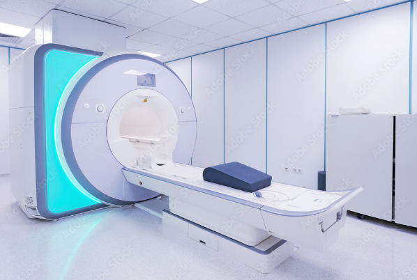
Advancements in MRI Technology
Recent advances in MRI technology have focused on improving imaging detail, speeding up scan times, and increasing patient comfort. These improvements allow clearer diagnosis and a broader range of clinical uses.
Key Innovations in Magnetic Resonance Imaging
New MRI systems feature larger bore sizes to reduce patient anxiety and accommodate a wider range of body types. High-field MRI machines now often operate at 3 Tesla or higher, increasing image clarity.
Functional MRI (fMRI) has grown in use, enabling doctors to observe brain activity by tracking blood flow changes. There are also novel MRI pulse sequences that allow for measuring tissue characteristics like fat content, fibrosis, and iron levels.
Innovations in hardware, like improved magnets and gradient coils, combine with advances in software to enhance the detail and scope of images captured.
Improvements in Image Resolution and Speed
Higher resolution MRI machines offer sharper and more detailed images. This helps detect small abnormalities earlier and more accurately.
Scan speeds have dramatically increased due to faster data processing and better algorithms. Faster scans reduce the time patients need to remain still, decreasing motion artifacts that blur images.
Innovations such as parallel imaging allow simultaneous data collection, further speeding exams without reducing image quality.
Enhanced Safety and Patient Comfort
Modern MRI systems focus on reducing noise, which can be loud and stressful. New designs and soundproofing minimize this discomfort during scans.
Non-invasive imaging means no radiation exposure, ensuring patient safety during repeated tests. Additionally, wider bores and shorter scan times help lessen claustrophobia.
Some MRI machines now include in-system entertainment to distract and relax patients, improving their overall experience and cooperation during scans.
Integration of Visual Technology with MRI
The combination of MRI with advanced visual technology improves how images are captured, viewed, and analyzed. This integration allows faster image updates, detailed 3D models, and the use of artificial intelligence to interpret complex data. Together, these elements provide clearer insights into patient conditions.
Real-Time Imaging and Data Visualization
MRI systems now support real-time imaging, which enables doctors to see changes inside the body as they happen. This capability is vital during surgeries or interventions, providing live feedback to guide decision-making.
Data visualization tools transform raw MRI scans into clear, color-coded images that highlight specific tissues or abnormalities. These visual aids help clinicians quickly identify key features without sifting through hundreds of raw slices.
Advanced graphical displays can combine different types of imaging data, such as MRI with CT scans, allowing for side-by-side or overlay views. This gives a more complete picture of the anatomy or pathology.
3D Reconstruction and Augmented Reality Applications
MRI data can be processed into detailed 3D models representing organs, blood vessels, or tumors. These models assist doctors in understanding complex anatomy from multiple angles before surgery or treatment.
Augmented reality (AR) uses these 3D reconstructions to project the images onto the patient or a screen during procedures. This helps surgeons visualize and navigate internal structures without making large incisions.
AR-assisted MRI guidance can reduce risks and improve precision in treatments, such as biopsies or radiation therapy. It also supports medical education by offering interactive, realistic anatomy views.
Artificial Intelligence in MRI Interpretation
Artificial intelligence (AI) systems analyze MRI data to detect patterns not easily seen by the human eye. AI can identify subtle changes in tissues, improving early diagnosis of diseases like cancer or brain disorders.
Machine learning algorithms enhance image quality and fuse multiple imaging types, such as PET and MRI, to provide more detailed information. AI can also speed up the interpretation process, delivering results faster.
By integrating AI, diagnostic accuracy improves, and workloads for radiologists decrease. This technology supports personalized treatment planning based on precise image-based insights.
Clinical Applications and Benefits
The fusion of MRI with advanced visual technology creates detailed images that help doctors see inside the body with clarity. This combination improves how diseases are detected, treated, and monitored. It also supports personalized care and better understanding of complex conditions.
Improved Diagnosis and Treatment Planning
Combining MRI with visual technology allows for clearer, more precise images of organs and tissues. This helps identify abnormalities early, such as tumors or vessel blockages. Clearer images reduce the chances of misdiagnosis and guide more accurate treatment plans.
Doctors can use these detailed images for planning surgeries or other treatments. For example, fusion imaging helps locate exact tumor boundaries or delicate brain areas to avoid during surgery. This precision lowers risks and can speed up patient recovery.
The real-time imaging offered by some fused MRI techniques adds value during procedures. It lets doctors adjust treatment immediately based on live feedback, making interventions safer and more effective.
Personalized Medicine and Patient Outcomes
Visual fusion with MRI supports tailoring treatments to each patient’s specific condition. By capturing detailed anatomy and function, doctors can design personalized care strategies. This approach improves treatment success rates and reduces unwanted side effects.
Better imaging allows continuous monitoring of how a patient responds to therapy. Adjustments can be made quickly if treatments are not effective. This dynamic process helps improve patient outcomes.
Patients also benefit from less invasive diagnostic methods. Fusion imaging often reduces the need for multiple separate scans or biopsies, lowering discomfort and risks.
Advanced Neurological and Musculoskeletal Imaging
In brain imaging, the fusion of MRI and visual technology maps both structure and brain activity. This helps identify critical regions to preserve during treatment and better understand neurological disorders.
For musculoskeletal issues, fused imaging reveals detailed views of bones, joints, and soft tissues. This helps detect injuries, inflammation, or degenerative changes earlier and with more accuracy.
Clinicians use these enhanced images to plan treatments that protect vital brain functions or optimize joint repair. This improves patient safety and treatment effectiveness in complex cases.
Future Trends and Emerging Challenges
The integration of MRI with visual technologies is advancing through new research, raising important ethical questions and data protection needs. Healthcare systems must also prepare for the effects of these innovations on costs, workflows, and patient care quality.
Ongoing Research and Developments
Current research focuses on improving MRI resolution and reducing scan times by combining AI with visual imaging techniques. Portable MRI devices with enhanced visuals are being developed to increase accessibility outside hospitals.
Photo-counting CT and whole-body MRI technologies are expanding, allowing more detailed and faster whole-body scans. AI-driven image analysis helps detect abnormalities earlier and with better accuracy.
Efforts to make machines eco-friendly and to include patient entertainment systems are underway. These improvements aim to enhance patient comfort and reduce MRI’s environmental impact while maintaining image precision.
Ethical Considerations and Data Security
The use of AI in MRI and visual fusion raises concerns about patient privacy and the security of imaging data. Strict protocols will be needed to prevent unauthorized access and misuse of sensitive health information.
Transparency about how AI algorithms operate is crucial to avoid biases that could affect diagnosis. Patients must be informed about automated decision-making processes linked to their scans.
Regulations will likely evolve to address these challenges, requiring healthcare providers and tech developers to work together closely. Ensuring data security will be a constant priority as imaging technologies become more integrated and connected.
Potential Impact on Healthcare Systems
These advances may drive higher upfront costs for hospitals due to new equipment and training needs. However, faster, more precise imaging can reduce repeat scans and improve treatment planning, potentially lowering long-term expenses.
Workflow changes will involve more collaboration between radiologists, AI specialists, and technicians. Tele-radiology and remote visual image analysis could expand service reach but require reliable internet and infrastructure.
Patient experience may improve through shorter scans and more transparent results, supporting shared decision-making. Still, healthcare systems must balance managing rising demand for imaging with maintaining affordable access.
Also Read :