Intraoperative MRI in Complex Neurosurgical Cases: Improving Precision and Outcomes
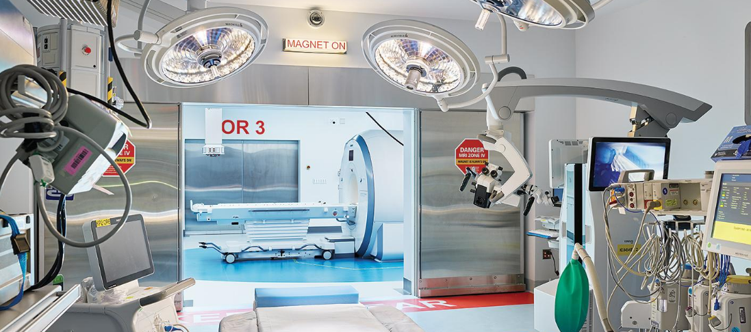
Intraoperative MRI is going to revolutionize the management of complex neurosurgical cases. Advanced technology provides surgeons with real-time brain images during surgery, thus enabling them to work with more precision. Intraoperative MRI enables surgeons to assess tumor removal intraoperatively and make immediate alterations to technique, which improves patient outcomes.
This technique is particularly vital when performing difficult procedures, such as brain or spine operations. Immediate feedback will reduce complications and allow surgeons to make informed decisions based on the most current information. This innovation enhances not only safety but also increases the chances of successful surgeries.
Neurosurgical intraoperative MRI is a very dynamic field. Its benefits weigh more and more due to clinical applications representing the milestones of neurosurgeons in very challenging cases.
Key Takeaways
- Intraoperative MRI offers imaging in real time during the surgery.
- It greatly enhances accuracy during the surgical procedure and improves patient outcomes.
- This technology is indispensable for complex neurosurgical procedures.
Technological Fundamentals of Intraoperative MRI
Intraoperative MRI is a combination of the most advanced imaging technique with a surgical procedure. It enhances the capability of visualization relating to brain structures by many folds in complex surgeries. This chapter addresses the development, core components, and differences between the standard and high-field systems.
Development and Evolution of Intraoperative MRI
The first intraoperative MRI systems came into the scene in the 1990s. These inventions were proposed for increasing the accuracy of brain surgery. Since then, the technology has significantly improved, evolving from big and bulky machines into slicker models.
Early systems took up long setup times and had limited imaging capabilities. Newer models can be moved more easily with high-resolution images provided often much faster. The integration of MRI during surgery allows real-time feedback to achieve better outcomes from surgery.
Modern systems also ensure safety and comfort for the patient. They minimize the usage of time in the operation room and maximize the quality of imaging. The continued research ensures these technologies will get better and further enhance surgical accuracy.
Core Components and Functional Principles
There are several main units that make up an intraoperative MRI. Some units that could be considered as most basic include the magnet, computer system, and imaging software.
The magnet provides the very strong magnetic field required for imaging. The protons in the body will actually align in a uniform manner and then emit signals. The computer will process these signals to create detailed images of the brain.
Such images will be visible to the surgeons with the help of the imaging software. This lets them identify the position of critical structures and make informed decisions based on the same. Advanced algorithms clear the images, and even small tumors or tender nerves become more visible.
Their efficiency is that they can be integrated with surgical tools. Surgeons can operate and, simultaneously, can keep on checking MRI images, which resulted in better precision during the operation.
Standard vs. High-Field Systems
Intraoperative MRI systems come with two prime variants-standard systems and high-field systems. Each of them has different applications and advantages.
Standard Systems:
Normally use 1.5 Tesla or a magnetic field of a lesser magnitude.
Smaller and more compact, hence easier to integrate into operating room theaters.
May be sufficient for the majority of surgical cases, although image detail is less compared to others.
High-Field Systems:
Operate at 3 Tesla or greater.
- Greater resolution and quality of images.
- Ideal for complicated surgeries or when minute details are important.
- Surgeons decide between these depending on the complexity of the case. High-field systems should be chosen when precision is of utmost importance, while for less intricate operations, standard ones will suffice.

Also Read :
